

In thirteen years an age-old anniversary of PAP-test, an effective and simple in usage method of revealing precancer and early carcinoma of uterine cervix, so called in honour of its creator, a Greek scientist Georgios Papanikolau, will be celebrated. Since 1943 the test has saved lots of women’s lives and it would have saved more if the innovation, suggested by doctor Papanikolau in 1928, had been objectively and in good time evaluated by doctor’s community instead of exposing to traditionally durable scepticism.
Nowadays the problem of screening precancerous and cancerous diseases of uterine body and appendages, which are increasing in population, is still unsolved. What do we know about it? Do we use the given diagnostic opportunities for preserving reproductive health and lives of our patients? It is the necessity of regular prophylactic measures, in the aspect of forming the considered diseases, that consists in high chances of their elimination just when they are early revealed.
As it is known, the problem of intensification and optimization of pre-hospital diagnostics largely determines the success of treatment and is one of its most important items. It is this approach that gives an opportunity for effective, preserving organs, treatment and thus at most saves reproductive abilities of women. It won’t be an exaggeration to say that early and, furthermore, differential diagnosing diseases of inner genitalia remains one of the most complicated problems of gynecology. It gains peculiar significance while forming the concept of revealing precancer, early forms of endometrial cancer and cancer of uterine appendages, when there are no symptoms or they are very scarce and sizes of pathological tumors are minimal, and are not spread on other organs (as it is known, in such cases, treatment is more sparing and effective, and maximally favourable). WHO experts opinion of the absence of reliable screening programs of precancerous and cancerous pathology of uterine body (endometrial cancer) and appendages should be also mentioned.
And, meanwhile, statistical data of Russian and foreign scientists convincingly testify not only steady increase of sick rate, but also ‟rejuvenation” of patients with benign and malignant hormone-dependent tumors of reproductive organs. This fact is explained not only by increase of an average life span, but also by the essential increase in population of such ‟diseases of civilization” as adiposity, anovulation, chronic hyperestrogenia, infertility, inflammatory diseases of uterine and appendages, and also by repeated ‟aggressive” obstetrical and gynecological manipulations [1, 2, 3]. That is why the necessity of forming a new approach to ‟active search” for patients with precancerous and early cancerous diseases of inner genitalia should be considered first of all from the position of elaborating the new screening test as one of the most perspective methods of their second prevention [4].
It is known that at cancerous growth mitochondrial oxygenation is forced out by glycolysis, more primitive way of energy supply. It is accompanied by active oxygen forms and is the basis of stimulating free radical oxygenation in the organism. During this process a state called «the oxydative stress» (OS) is developed and results in accumulating the highly toxic combinations leading to morphological disturbances of cells and tissues [5, 6, 7].
Free radicals hit all kinds of biological macromolecules, furthermore, the most studied are processes of lipids peroxide oxygenation. However, only active forms of oxygen also cause oxygenative destruction of proteins. It is considered that in a state of OS lipids are not attacked by the active oxygen forms first and foremost, but proteins of cells and biological fluids (BF) are. This process may also be the basic in structural and functional disturbances of the reproductive system organs, including the development of precancerous and cancerous transformation of endometrial and ovarian tissues. However, it needs a considerable amount of pathologic deformation of molecular ingredients of a cell and, consequently, a prolonged time interval, in the final stage of which traditional cytologic and histologic methods of research are already available. In it lies one of the reasons of the displacement of the forming clinical picture and, consequently, the initiation of diagnostic measures of the disease for a later period.
It is important to note that structural changes of endometrium (E), as a hormone-dependent tissue, largely depend on the functional condition of ovaria. It is the endometrial tissue that reacts most sensitively upon the changes of homeostasis of the reproductive system organs, also including functional disturbances while forming tumors of gonades. According to it, we should point out the ability of a number of enzymes and their isoforms to accumulate during hyperplastic processes and cancer of endometrium in the apical parts of glandular cells, and then to secrete into the endometrial secretion [8]. It should be noted that besides blood and fragments of sloughing endometrial tissue, endometrial and endocervical secretion is also included into menstrual fluid (MF). By the drawn analogy the OS markers, among which the earliest is the carbonyl group of proteins (CGP), may also be found with the help of simple methods in MF and endometrial lavages (EL) of examined patients long before appearing manifesting symptoms of the disease.
Choosing MF (and EL), as an object of research, is explained not only by scientific curiosity (because they are poorly studied!), but also by the fact that once a month this BF in enough volume for examining (from 10 to 80 ml in healthy women) is spontaneously secreted from the uterine cavity and can be collected by patients themselves (into sanitary napkins or special caps) for the laboratory examination.
Traditionally morphological research in medicine is mainly concentrated on tissues and cellular elements whilst structural peculiarities of BF remain poorly studied. In this respect microscopy of the structures of a dry drop (facies), forming in BF when it turns into a solid state as a result of dehydration, is long-range. This new scientific trend was called «morphology of BF» [2]. The essence of the new method of diagnostics is in the fact that normally the microscopic picture of the examined drop of BF has more or less homogeneous structure and by slight influence of different damaging factors (oxidizing, toxic) the hydrate membranes of proteins change, and processes of molecular aggregation start.
In professors V.N. Shabalina and S.N. Shatohina opinion (1995, 2007), facies (F) is a structural portrait of molecular correlation in BF which allows to put deep microprocesses on the macrolevel. A microscopic picture of F has structures which are characteristic for every kind of human BF, connected with peculiarities of physiologic and pathologic processes action in the organism.
Using technologies of the morphological analysis «Lithos-system» [10] one can observe even the earliest pathologic changes of molecular structures immediately, without «incubation period», which is necessary at cellular, organic and/or systemic levels. Therefore, the morphologic analysis of BF gives an opportunity for the earliest diagnostics of the developing pathologic process.
We offer a quantitative content analysis of the OS markers with a simultaneous defining structural microscopic peculiarities of the solid phase of MF and EL as a new diagnostic (and prognostic) approach to evaluating the state of inner genitalia in women of reproductive and (especially) perimenopausal ages.
The inner anatomical localization, complexity of the microscopic structures of neoplasms of the female gonades, indistinct borders between the neoplasms of a different maturation degree and diversity of their combinations – all these complicate the early revealing of ovarian tumors (OT) and hinder rational planning for patients treatment.
As it was already told, E tissue is extremely sensitive to the homeostatic changes of the reproductive system organs also including the increase of the OS markers content in EL, in particular, CGP by forming OT. Thus, according to D.L. Ovodenko et al. (2008), women from the control group with the normal ovarian structure had 0–1,9 (0,9 ± 0,14) nmol/mg. While increasing the index up to 2,0–3,5 (2,8 ± 0,28) nmol/mg in the majority of cases there were revealed benign tumors (p < 0,05) and at 3,6–4,9 (4,8 ± 0,25) nmol/mg – malignant OT (p < 0,01).
The CGP content in the MF of the patients with the benign ovarian tumors (BOT) was higher than the patients with chronic inflammatory processes of uterine appendages (CIPUA) and those from the control group had (p < 0,01).
The microscopic picture of F of EL in the women from the control group was characterized by sharply shaped zone borders. In the majority of cases there were two zones – peripheral with radial cracks and central, containing crystals of salts, more rarely – straight radial cracks as a prolongation of the peripheral zone cracks (Fig. 1). Besides that, in 24,8 % of the cases in F of EL of the women from this group appeared the third intermediate zone, located between the two above mentioned zones, containing three-rayed cracks.
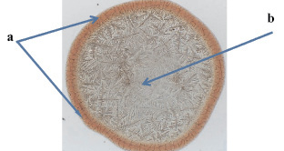
Fig. 1. Facies of endometrial lavage of reference group patient (×10) а – peripheral zone with radial cracks; b – salt area
In the patients with CIPUA and during the aggravation of the process, the area sizes of three-rayed and radial cracks didn’t reliably differ from the analogous indexes of the women from the control group. However, in these cases «tongue» structures (markers of inflammation), «twirls» and wisp blocks (markers of hypoxia) appeared in the picture of F.
F of EL in the patients with benign ovarian tumors and ovarian cancer were rather different in their structure from those, which the women from the control group had. Facies zones of EL remained in the patients with benign ovarian tumors of all age groups, moreover, in 68,4 % of cases they have already had the zone with three-rayed cracks (Fig. 2).
In those patients, who had ovarian cancer, the area of the three-rayed cracks zone on the surface of F markedly enlarged up to 2,2 ± 0,10 mm2 (Fig. 3), being reliably different from the same in the control group (p < 0,01) and in the group of patients with benign ovarian tumors (p < 0,05). The area of the peripheral zone with radial cracks was 6,1 ± 0,42 mm2 and in size it wasn’t different from that, which the women from the control group had [11].
The obtained results testify that the microscopic picture of F of EL and MF has clear and distinctive peculiarities in patients with benign, frontier and malignant ovarian tumors. Including methods of structural and biochemical analysis of EL and MF into a diagnostic complex in the patients from “risk groups” of the OT development will allow to increase sensibility and to rise specificity of the research [12].
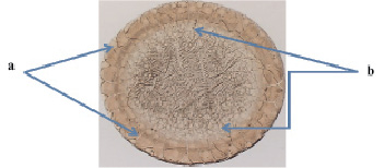
Fig.2. Facies of endometrial lavage of patient with benign OT, (×10) а – peripheral zone with radial cracks; b – three-beam cracks
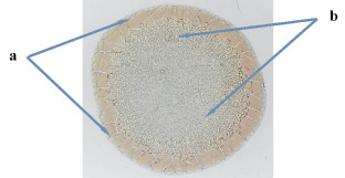
Fig. 3. Facies of endometrial lavage of patient with malignant OT, (×10) a – peripheral zone with radial cracks; b – three-beam cracks
An important advantage of the offered method of diagnostics is noninvasive and autraumatic collecting of the researched material, the possibility of multiple repeating the research in antenatal clinics, making analysis in laboratories of Health institutions. The obtained results confirm the possibility of revealing ovarian cancer at early stages of its development when organ-preserving surgery becomes the most realistic and, thus preserving reproductive health of women.
Библиографическая ссылка
Shvarev E.G., Dikareva L.V., Ovodenko D.L., Aupova A.K., Shvarev G.E. An Innovative Approach of Adnexis Tumors Diagnostics // Международный журнал экспериментального образования. 2015. № 12-6. С. 729-732;URL: https://expeducation.ru/ru/article/view?id=9325 (дата обращения: 08.07.2026).